

In modern medicine, accurate diagnosis is the absolute prerequisite for effective treatment. At the core of this diagnostic power is the Radiology Department (Medical Imaging). Over the past two decades, imaging technology has evolved from simple film X-Rays to highly advanced, AI-assisted digital modalities that can construct 3D models of internal organs in seconds.
Establishing a radiology department is one of the most capital-intensive and architecturally demanding projects a healthcare facility can undertake. The equipment is heavy, highly sensitive, and emits varying types of radiation or magnetic fields that require specialized room construction. In this guide, Mak Medikal details the essential equipment and the critical infrastructure required to build a cutting-edge radiology department.
A comprehensive radiology department must be equipped to handle everything from bone fractures to complex neurological diagnostics. Here are the foundational devices:
The backbone of any imaging center. Modern systems have completely moved away from chemical film processing to Digital Radiography (DR). DR systems use flat-panel detectors that capture X-rays and instantly convert them into digital images displayed on a monitor. This drastically reduces radiation exposure to the patient, increases patient throughput, and allows images to be enhanced and shared instantly across the hospital network.
A CT scanner takes hundreds of cross-sectional X-ray images as it rotates around the patient, which a computer then compiles into detailed 3D images of bones, blood vessels, and soft tissues. When selecting a CT scanner, the "slice count" (e.g., 16-slice, 64-slice, 128-slice) is crucial. A 64-slice or 128-slice CT is the gold standard for cardiac imaging and advanced trauma cases, offering incredibly fast scan times that reduce motion artifacts.
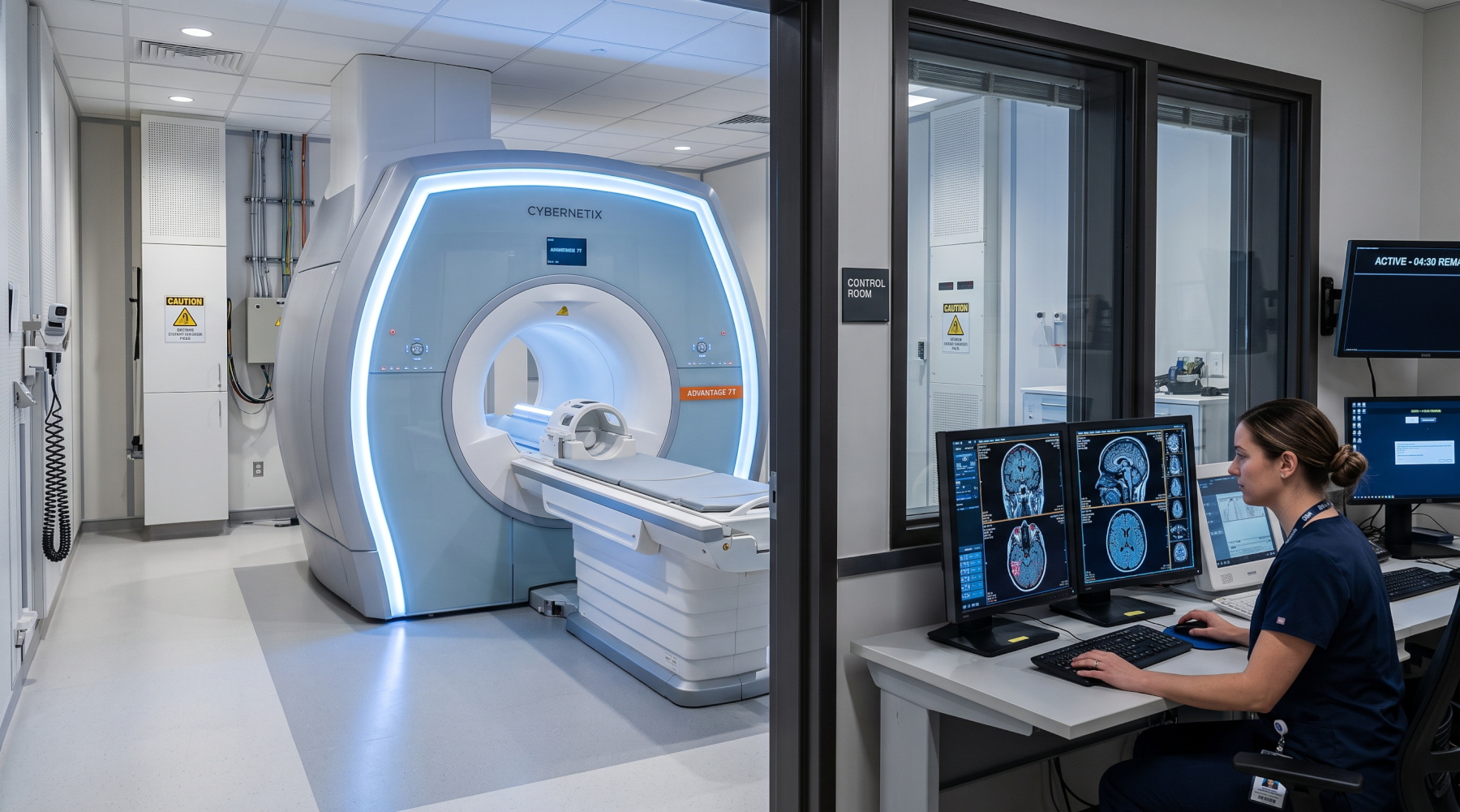
Unlike X-Rays and CTs, MRI does not use ionizing radiation. Instead, it uses powerful magnets and radio waves to create exceptionally detailed images, particularly of the brain, spinal cord, ligaments, and soft tissues. MRIs are categorized by magnetic field strength, measured in Tesla (T). A 1.5T MRI is the industry workhorse for general imaging, while a 3.0T MRI offers ultra-high resolution required for advanced neurology and research.
Ultrasound uses high-frequency sound waves to capture live images. It is highly versatile, radiation-free, and essential for obstetrics/gynecology, cardiology (Echocardiography), and internal medicine. A modern department needs a mix of high-end cart-based ultrasound machines with various probes (convex, linear, phased array) and portable units for use in the ICU or Emergency Room.
Fluoroscopy provides continuous, real-time X-ray imaging, functioning like an "X-ray movie." It is vital for gastrointestinal studies and angiography. In the operating room, mobile versions called C-Arms are indispensable for guiding orthopedic surgeons during bone fixations or assisting in vascular stent placements.
You cannot simply plug an MRI or CT scanner into a standard room. The architectural demands of a radiology department are immense.
Radiation Shielding (Lead Lining): Rooms housing X-Ray, CT, and Fluoroscopy machines must have lead-lined walls, doors, and viewing windows to protect hospital staff and the public from ionizing radiation scatter. The thickness of the lead is calculated by medical physicists based on the machine's power and room size.
Faraday Cages for MRI: An MRI machine generates a magnetic field thousands of times stronger than the earth's magnetic field. The MRI room must be enclosed in a Faraday Cage (usually a copper-lined room) to prevent external radio frequencies (like FM radio or cell signals) from interfering with the image, and to contain the magnetic field within the room.
Structural Reinforcement and Cooling: MRI and CT machines weigh several tons; floor loading capacities must be reinforced. Furthermore, these machines generate massive amounts of heat. Dedicated, heavy-duty chilling systems (chillers) and specialized HVAC units are required to keep the equipment from overheating.
A modern radiology department is completely digital and film-less. This requires powerful software architecture:
PACS (Picture Archiving and Communication System): A specialized server network that securely stores digital imaging files (in DICOM format) and allows doctors to access and view them on high-definition diagnostic monitors anywhere in the hospital.
RIS (Radiology Information System): The software used to manage patient scheduling, billing, and radiological reporting, integrating seamlessly with the hospital's main Information System (HIS).
Building a radiology department requires millions of dollars in CAPEX. Mak Medikal offers a strategic solution to optimize this budget: Certified Refurbished Imaging Systems.
Purchasing a refurbished 1.5T MRI or a 64-slice CT scanner from top global brands through Mak Medikal can save your facility up to 50% of the cost of a new machine. These systems undergo rigorous factory-level reconditioning, tube replacements, and software upgrades, coming with comprehensive warranties and maintenance contracts. This allows clinics in emerging markets to offer world-class diagnostics at a fraction of the initial investment.
Mak Medikal provides end-to-end radiology solutions—from the initial lead-shielding calculations and architectural drawings to the supply, installation, and software integration of the most advanced imaging modalities on the market.
A comprehensive guide to setting up a hospital radiology department. Discover key imaging devices like MRI, CT, X-Ray, Ultrasound, and infrastructure needs.
Explore the ultimate guide to Operating Room (OR) equipment. Learn about surgical tables, anesthesia machines, lighting, and turnkey OR setup solutions.
Discover why Turkey is the leading hub for medical equipment export. Explore turnkey hospital solutions, logistics to Africa & MENA, and top device categories.